Advancing Vision Care with Cutting-Edge Solutions
How to diagnose glaucoma?
Measuring the pressure in the eye is only one of many tests required to confirm the diagnosis. Other tests include examining the drainage angle, measuring the corneal thickness, testing the visual field and scanning the optic nerve are also important.
Some of these tests need to be repeated periodically to monitor the progression of the disease.
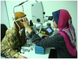
Laser treatment for glaucoma
Screening for Glaucoma
Age 20-29 years
- at least once during this period
- people with family history of glaucoma, diabetes, previous serious eye injury or nearsightedness should be screened every 3-5 years
Age 30-39 years
- at least twice during this period
- those with risk factors (as above) should be seen every 2-4 years
Age 40-64 years
- every 2 years
Age 65 years or older
- every year
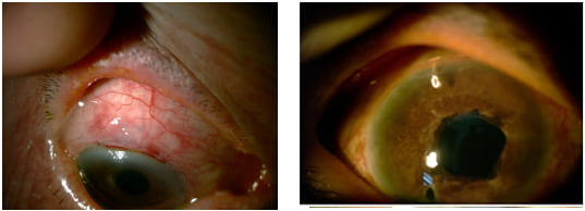
Formation of a ‘bleb’ after trabeculectomy and Optonol Shunt – a metal tube implant
Frequently Asked Questions (FAQs)
Answers to Your Frequently Asked Questions: Clearing Up Common Queries
Glaucoma is the second leading cause of blindness worldwide and is characterized by raised pressure inside the eye which causes damage to the optic nerve leading to permanent loss of vision.
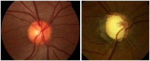
Normal optic nerve head(left) and damaged optic nerve head from glaucoma (right)

Tunnel vision with end stage glaucoma (right)
- Occurs most frequently in people over 60 years but can happen at any age.
- Association with race – Asians are more prone to closed angle glaucoma whereas Afro-Caribbeans tend to get open angle glaucoma
- Diabetics are more prone to developing glaucoma
- People who are nearsighted are more prone to open angle glaucoma whereas those who are farsighted tend to get closed angle glaucoma
- Serious eye injury can sometimes lead to glaucoma
- Steroid medication can also cause glaucoma when it is used for a long time
- Glaucoma can sometimes be inherited in certain families
Open angle glaucoma is the more common and occurs when there is gradual build up of pressure within the eye even when the drainage angle is not blocked. Visual loss occurs gradually and often goes unnoticed by the patient until it is quite advanced.
Closed angle glaucoma occurs when there is physical blockage of the drainage angle and there can sometimes be a sudden build up of pressure associated with pain, headache, nausea and vomiting.
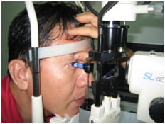
Measuring intraocular pressure
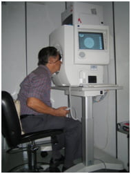
Visual field assessment
When glaucoma is advanced, surgery may be required to lower the eye pressure and Minimally Invasive Glaucoma Surgery (MIGS) is the latest technology available whereby microstents (or tubes) are inserted into the eye to improve drainage of fluid and lower pressure.
More Related Topics
Children’s Eye Disorders

Retinal Detachment

Retinal Diseases & Vitrectomy

Contact Lenses & Glasses

Retinal Diseases & Vitrectomy

Children’s Eye Disorders

Retinal Detachment

Contact Lenses & Glasses
Get In Touch
If you are interested in talking to us about our clinical services, please send us a message.

