As diabetes is a systemic disease, optimal control of blood sugar as well as blood pressure and cholesterol levels are still very important in the treatment of diabetic retinopathy. However, progression of retinopathy may occur despite all medical efforts. If diabetic retinopathy is detected early, treatment with laser photocoagulation may stop continued damage. Even in the advanced stages of the disease, laser treatment can reduce the chance that a patient will have severe visual loss.
Laser treatment is used to seal or obliterate the abnormal leaking blood vessels. This procedure focuses a powerful beam of laser light onto the damaged retina. Small bursts of the laser energy seal leaking vessels and form tiny scars inside the eye. The scars reduce new vessel growth and cause existing ones to shrink and close. Laser treatments are usually carried out in an outpatient setting.
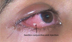
Eye redness and watering from conjunctivitis
Allergies affecting the eye are fairly common. The most common allergies are those related to pollen, particularly when the weather is warm and dry. Symptoms can include redness, itching, tearing, burning, stinging, and watery discharge, although they are not usually severe enough to require medical attention.
An increasing number of eye allergy cases are related to medications and contact lens wear. Also, animal hair and certain cosmetics, such as mascara, face creams, and eyebrow pencil, can cause allergies that affect the eye. Touching or rubbing eyes after handling nail polish, soaps, or chemicals may cause an allergic reaction. Some people have sensitivity to lip gloss and eye makeup. Allergy symptoms are temporary and can be eliminated by not having contact with the offending cosmetic or detergent.
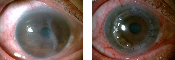
Corneal Infections. Sometimes the cornea is damaged after a foreign object has penetrated the tissue, such as from a poke in the eye. At other times, bacteria or fungi from a contaminated contact lens can pass into the cornea. Situations like these can cause painful inflammation and corneal infections called keratitis. These infections can reduce visual clarity, produce corneal discharges, and perhaps erode the cornea. Corneal infections can also lead to corneal scarring, which can impair vision and may require a corneal transplant.
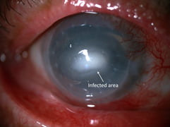
Infection of the cornea with diffuse eye redness and inflammation
As a general rule, the deeper the corneal infection, the more severe the symptoms and complications. It should be noted that corneal infections, although relatively infrequent, are the most serious complication of contact lens wear.
Infection of the cornea with diffuse eye redness and inflammation
Dry Eye. The continuous production and drainage of tears is important to the eye’s health. Tears keep the eye moist, help wounds heal, and protect against eye infection. In people with dry eye, the eye produces fewer or less quality tears and is unable to keep its surface lubricated and comfortable.As we age, the eyes usually produce fewer tears. Also, in some cases, the lipid and mucus layers produced by the eye are of such poor quality that tears cannot remain in the eye long enough to keep the eye sufficiently lubricated. The main symptom of dry eye is usually a scratchy or sandy feeling as if something is in the eye. Other symptoms may include stinging or burning of the eye; episodes of excess tearing that follow periods of very dry sensation; a stringy discharge from the eye; pain and redness of the eye. Sometimes people with dry eye experience heaviness of the eyelids or blurred, changing, or decreased vision, although loss of vision is uncommon.