LEC Eye Centre Sdn. Bhd.

Specialist Eye Care Centre

Age-Related Macular Degeneration

Advanced Clinical Services for Age-Related Macular Degeneration

Age-Related Macular Degeneration

Defying Age, Defending Sight

Age-Related Macular Degeneration

Defying Age, Defending Sight

How to detect AMD?

AMD only affects the central vision, hence, symptoms of disease can range from mild central blurring to distortion of images or a complete ‘blind spot’ in the centre of vision.

An effective way of detecting AMD is through regular eye checkups with your ophthalmologist particularly if there are associated risk factors. There are also sophisticated investigations which can be carried out to evaluate the extent of disease involvement and these tests include Fundus angiography where a dye is injected into a vein in the arm, and photographs of the retina are taken as the dye is carried in the blood stream to the eye.

Another very detailed scan of the eye called Optical Coherent Tomography can be done as well.

Colour photo of macula (top left), FFA image (top right) and OCT (bottom)

Treatment Of AMD

Dry AMD – In advanced dry AMD, there is currently no form of treatment which can prevent visual loss. However, in the earlier forms of dry AMD, there is strong evidence that taking a oral formulation of anti-oxidants and zinc can reduce the risk of progression to advanced AMD.

Wet AMD – In the past, treatments available could only slow down the process of visual loss in this disease. These treatments included laser surgery and photodynamic therapy. More recently, there have been a development of a new group of drugs for injection into the eye.

These group of drugs are known as anti-VEGF agents (Vascular Endothelial Growth Factor). From large clinical trials, it has been shown that this growth factor is in abundance in wet AMD, and regular injections of anti-VEGF agents can prevent significant visual loss in the majority of patients and even improve vision in up to a third of them.

Commonly used anti-VEGF drugs

Retinal surgery

Vitrectomy is a specialized microsurgical procedure which is used to treat retinal disorders.

The surgeon uses an operating microscope with a specialized viewing system to allow for appropriate magnification and a highly sophisticated vitrectomy machine together with very fine instruments to perform the surgery.

This is usually performed under local anaesthesia and is often an ambulatory procedure.The name vitrectomy refers to the removal of vitreous. Vitreous is a jelly-like structure which fills the back cavity of the eye. It is firmly attached to the retina in early life and with age, the vitreous changes to a watery form and starts to detach from the retina.

Consequently, clumps of jelly can form and float around casting shadows on the retina perceived as “floaters”. This is usually benign but it is often the bane of vitreoretinal surgeons as in certain diseases such as retinal detachment and proliferative diabetic retinopathy, the vitreous is the main culprit in the disease process causing significant visual disability if left unchecked.

The initial step in vitrectomy is to make 3 separate entry wounds into the eye. (Figure 3) The wound sizes are very small, ranging from 0.5mm to 1mm and can seal without sutures. The vitreous is removed using a miniature handheld cutting device and during the operation, the eye is refilled with a solution which is similar to what is being removed. A high intensity fibreoptic light is used to illuminate the back of the eye as the surgeon works.

Vitrectomy in progress(left), 3 openings into the eye for infusion, light source and vitreous cutter(top right) and surgeon’s view during surgery(bottom right)

Frequently Asked Questions (FAQs)

Answers to Your Frequently Asked Questions: Clearing Up Common Queries

AMD is a disease which affects the macula which is the part of the retina responsible for 90% of our functional vision. AMD usually affects individuals older than 55 years of age and causes blurring of the central vision which is required for activities such as reading and driving. AMD can be divided into 2 broad categories – dry and wet AMD.


Figure 1: Normal vision (left) and vision with AMD (right)

The dry form of AMD is usually more slowly progressive and affects approximately 90% of individuals with AMD. There is a gradual degeneration and loss of cells in the macula resulting in blurring of central vision. The most common sign of dry AMD is the presence of drusen, which are yellowish deposits under the retina. Dry AMD can range from mild to intermediate to advance depending on the extent of cell loss and the amount of drusen present. The more advanced the stage of AMD, the more significant the central visual loss.
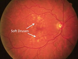
 

Figure 2: Dry AMD

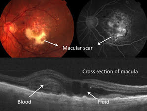
The wet form of AMD affects only 10% of individuals with AMD but is responsible for the majority of visual loss in this disease. There is a growth of a network of abnormal blood vessels under the retina and when these bleed, there is a sudden loss of central vision. The blood does eventually get reabsorbed but leaving behind permanent scarring and irreversible central vision loss.

A subset of wet AMD known as Polypoidal Choroidal Vasculopathy(PCV) seems to be more common in our Asian population. There is also bleeding from abnormal vessels under the retina but in this case, the vessels can be found in clusters or ‘polyps’.

Both these types of AMD often co-exist. Patients who have wet AMD will invariably have signs of dry AMD as well. However, patients with dry AMD do not always progress to develop the wet form of the disease and there is no way of telling which patients will progress and which would not.


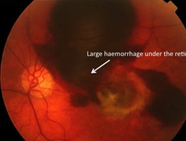
Figure 3: Sub-retinal bleeding in wet AMD

  1. Age – AMD tends to affect individuals above the age of 55 years old and the risk of developing this disease increases with increasing age.
  2. Smoking – There is evidence from large clinical studies which suggests a strong link between smoking and the development of AMD
  3. Family history – There is also strong evidence from studies suggesting a genetic component in this disease. Several genes have been found to be strongly associated with the development of AMD.

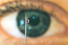
Gruesome as it may sound, this does involve “sticking a needle” into the eye and releasing specific drugs into the vitreous cavity. However, it does not hurt as much as most people think and it literally takes seconds to perform. It is used for the treatment of a variety of eye diseases but in recent years, it has revolutionized the management of AMD and has a significant impact not just on affected patients but also on retinal surgeons’ lives!!

The rise of intravitreal injections started in 2004 when intravitreal Pegaptanib sodium injection was first used in the management of AMD. This drug was developed to inhibit vascular endothelial growth factor (VEGF) which is a growth factor involved in the development of blood vessels. This had modest effect on the disease but the following year when Avastin (another inhibitor of VEGF) was first used to treat AMD, the revolution truly took off!

The impact of this ‘revolution’ extended beyond its positive effects on patients but also the increased workload for retinal surgeons as these injections had to be given every month in order to maintain its efficacy. There were also far reaching consequences for healthcare services in some countries as reimbursement for this very expensive drug burned very large holes in the healthcare budget!

Prevention better than cure

  1. If you have AMD, you should have a complete eye examination at least once a year by your eye specialist.
  2. If you have a family history of AMD, regular eye checkups also plays an important role
  3. Your lifestyle can impact on your risk of developing AMD:-
  • Don’t smoke
  • Have a healthy diet – green leafy vegetables and fish
  • Watch your weight
  • Exercise regularly

More Related Topics

Cataract

Glaucoma

Diabetic Retinopathy

Retinal Diseases & Vitrectomy

Cataract

Glaucoma

Diabetic Retinopathy

Retinal Diseases & Vitrectomy

Get In Touch

If you are interested in talking to us about our clinical services, please send us a message.